


 Innovation Highlights

3D Histology Technology-VIVIT Series
-
High-Fidelity Transparency with Minimal DistortionLess than 1% deformation ensures subcellular structures remain intact.Less than 1% deformation ensures subcellular structures remain intact.
Experimental data show that whole-brain samples treated with VIVIT exhibit minimal dimensional changes (–0.5% in length and –0.1% in width). The reagent has been validated across complex tissues including brain, multiple organs, and tumor models, and preserves subcellular structures such as synapses, providing a solid basis for 3D reconstruction and follow-up analysis. -
Signal Enhancement: Suppressing Photobleaching and Capturing Weak SignalsUp to 37-fold fluorescence enhancement, with markedly reduced background fluorescence.VIVIT can optimize the fluorescent environment after tissue clearing. Its ionic liquid formulation enhances both protein- and dye-based fluorescence while suppressing bleaching, quenching, and diffusion, therefore lowering background fluorescence.
Experimental results show that VIVIT increases protein-based signals by 2–3 times and amplifies dye-based fluorescence even more substantially, with Cy3 enhanced up to 37-fold. By maintaining dye quantum efficiency, VIVIT improves the ratio of target signal to background fluorescence and ensures continuity, preventing discontinuities such as stitching artifacts during 3D reconstruction. -
Cryo-Stability: Crystal-Free Freezing for Long-Term ReuseWithstands at least three freeze–thaw cycles and remains stable for imaging after long-term storage.VIVIT uses a crystal-free vitrification approach that prevents the structural damage and signal loss typically caused by ice-crystal formation during freezing, while enabling long-term preservation at –80 °C.
Experimental results show that VIVIT-treated samples withstand at least three freeze–thaw cycles without visible damage and remain suitable for direct 3D imaging even after 13 months of continuous storage. This performance far surpasses PBS or sucrose-embedded tissues, making VIVIT ideal for multi-round imaging and large-scale analysis of valuable samples.
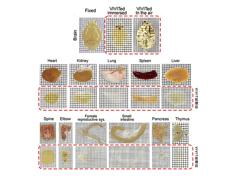
High-Fidelity Transparency with Minimal Distortion
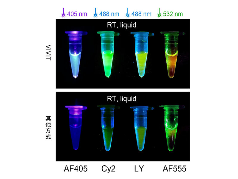
Signal Enhancement: Suppressing Photobleaching and Capturing Weak Signals
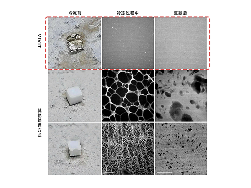
Cryo-Stability: Crystal-Free Freezing for Long-Term Reuse
3D Labeling Technology - RGB & LTD
Simultaneous Multi-Modal Labeling:
Presenting a Comprehensive View in One Sample
Compatible with diverse staining protocols, supporting multi-round labeling and imaging
TP MedTech’s 3D labeling system includes non-specific and specific staining approaches. It enables multi-modal and multi-round labeling of carbohydrates, nucleic acids, proteins, and other targets within the same sample.
-
RGB Non-Specific StainingProvides rapid three-channel staining without disrupting tissue integrity, enabling 3D-compatible H&E- and PAS-like protocols that present cellular and tissue morphology.
-
LTD Specific StainingUses directly conjugated antibodies together with penetration-enhancing mechanisms to achieve one-round multi-color labeling. The panel currently covers 22 markers, including immune, nucleic acid, glial, and neuronal targets, with stable signals and high antibody reusability.
RGB Non-Specific Staining
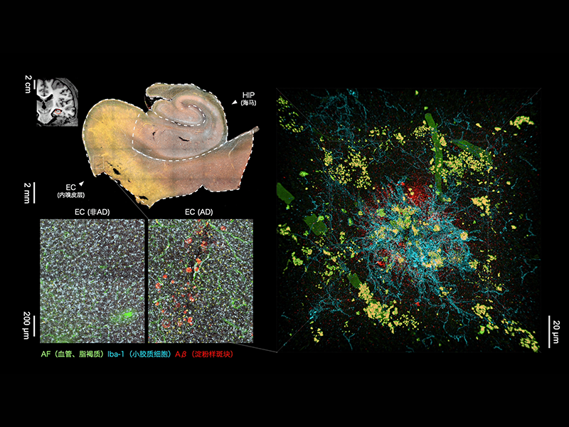
LTD Specific Staining
3D Reconstruction Technology – TARRS
3D Reconstruction: Millimeter-Scale Sections with Continuous and Accurate Stitching
Proprietary algorithm achieves subcellular-level registration with micron-level accuracy
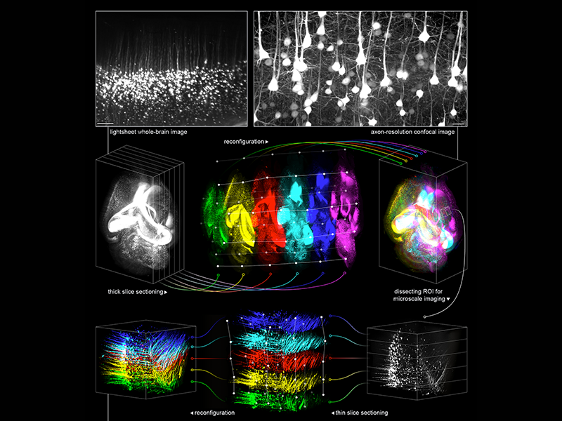
The proprietary TARRS algorithm enables subcellular-level 3D registration and reconstruction of millimeter-scale thick sections, with accuracy controlled at the micron level.
Building on VIVIT’s innovations in tissue clearing, which prevent ice-crystal damage and minimize tissue deformation, TARRS reduces discontinuities and distortions. This enables whole-brain reconstruction with only 2–3 thick sections, instead of the tens of thousands of thin sections required by conventional methods, improving both efficiency and accuracy.
Building on VIVIT’s innovations in tissue clearing, which prevent ice-crystal damage and minimize tissue deformation, TARRS reduces discontinuities and distortions. This enables whole-brain reconstruction with only 2–3 thick sections, instead of the tens of thousands of thin sections required by conventional methods, improving both efficiency and accuracy.
Whole-Brain 3D Reconstruction with VIVIT
3D-AI Modeling
AI Modeling: 3D Datasets Driving Intelligent Insights
High-content and multi-modal 3D spatial data enabling spatial modeling and intelligent diagnosis
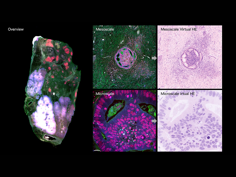
Our 3D spatial multi-omics platform provides high-content, multi-modal datasets from thick tissue sections, with each offering over ten-thousand times the information of a conventional 2D slide.
Building on these datasets, TP MedTech has developed a 3D-AI platform with modules for feature extraction and structural annotation. The model can process structural recognition, modeling, and prediction with only a minimal number of sections, and has been validated in gliomas, lung cancer, and lymphomas. It supports cases such as spatial subtyping, recurrence prediction, and therapeutic evaluation.
Building on these datasets, TP MedTech has developed a 3D-AI platform with modules for feature extraction and structural annotation. The model can process structural recognition, modeling, and prediction with only a minimal number of sections, and has been validated in gliomas, lung cancer, and lymphomas. It supports cases such as spatial subtyping, recurrence prediction, and therapeutic evaluation.
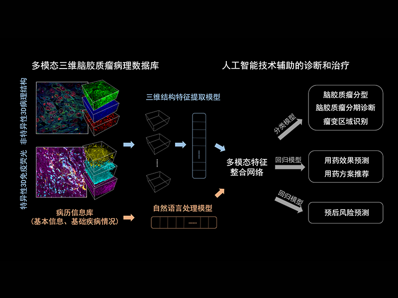
AI-Driven Diagnosis and Treatment with a Multimodal 3D Glioma Case Database
From Sample Processing to Data Analysis:
An Integrated 3D Spatial Multi-Omics Platform
By connecting the full workflow from tissue processing and molecular labeling, to image reconstruction and AI modeling, TP MedTech has built an integrated platform for 3D spatial multi-omics.
Through proprietary reagents, instruments, and AI models, we have achieved breakthroughs in structural fidelity, signal enhancement, thick-section reconstruction, and intelligent analysis, providing a solid foundation of 3D spatial data for basic research, clinical diagnosis, and drug development.
Through proprietary reagents, instruments, and AI models, we have achieved breakthroughs in structural fidelity, signal enhancement, thick-section reconstruction, and intelligent analysis, providing a solid foundation of 3D spatial data for basic research, clinical diagnosis, and drug development.